sydney neurosurgeon professor brian owler

Cervical Spinal Canal Stenosis & Myelopathy

What parts does the cervical spine comprise of?

The cervical spine is the upper part of the spinal column and is made up of seven vertebrae (C1–C7). These are the smallest vertebrae in the spine and support the weight of the head. The first vertebra, known as the atlas (C1), supports the skull, while the second vertebra, called the axis (C2), enables the head to rotate. Between most vertebrae are discs that cushion the spine and allow movement; however, there is no disc between C1 and C2.

Behind the vertebral bodies is a canal formed by a ring of bone that houses the spinal cord as it travels from the brain into the rest of the body. The spinal cord passes through this space and gives rise to nerve roots that exit the spine through openings known as intervertebral foramina. These nerve roots provide sensation and muscle control to the shoulders, arms, and parts of the upper back. The back of the vertebral ring is formed by the lamina and spinous processes, which act as attachment points for muscles and ligaments that support the head and neck.

Each disc in the cervical spine has two main parts: the annulus fibrosus, which is the tough outer layer of fibres, and the nucleus pulposus, the softer, gel-like centre that provides cushioning. Together, these structures allow flexibility, protect the spinal cord and nerves, and maintain stability of the neck during movement.

What is a cervical spinal canal stenosis

What is a cervical spinal canal stenosis?

The spinal cord which carries the messages from the brain to the structures of the body and vice versa runs though the spinal canal. The spinal canal is a tube of bony vertebrae with ligaments and discs between each vertebra.

Stenosis refers to a narrowing of the spinal canal and therefore cervical spinal canal stenosis results in compression of the cervical spinal cord.

In most cases this a degenerative process that occurs over a long period due to a combination of disc bulging from the front of the spine back towards the spinal cord, and buckling of the ligaments running between the bones at the back of the spine. The combination of the bulging discs and buckling ligaments pinches or compresses the spinal cord, sometimes over multiple levels.

There are other causes of cervical canal stenosis such as ossification of the posterior longitudinal ligament (OPLL), which is common in people of some ethnicities. In addition, movement of one bone on another, or malalignment of the cervical spine can also lead to compression of the cervical spinal cord.

What is cervical myelopathy?

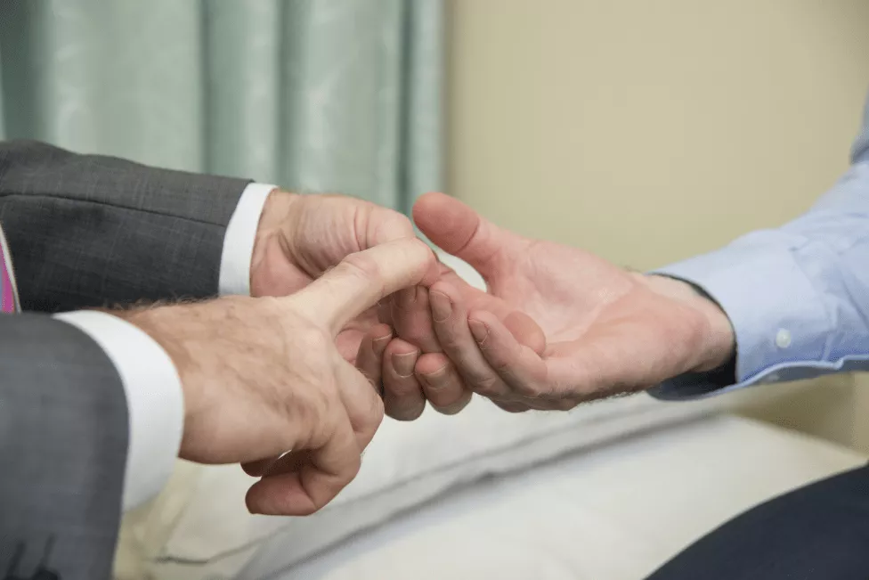
Cervical myelopathy is a clinical condition due to cervical canal stenosis. It is characterised by stiffness and clumsiness of the hands which are often numb. Commonly patients complain of a sandpaper like feeling in their hands. Walking and balance (gait) may also be affected, such that people may stumble easily and frequently fall.

Examination of a patient will often demonstrate weakness of the upper limbs and hands. There may be wasting of the small muscles within the hands themselves. The reflexes in the upper and lower limbs may be increased rather than depressed. The signs may be subtle in the early stages.

However, for most patients, there is no, or minimal neck pain associated with cervical myelopathy. Therefore, patients will often present to their doctor in a delayed fashion or diagnosis may be delayed.

Early recognition and referral are important to prevent deterioration that can be difficult to reverse. MRI of the cervical spine will confirm whether there is cervical canal stenosis and whether the spinal cord is compressed.

How is cervical canal stenosis and cervical myelopathy treated?

The treatment for cervical canal stenosis and/or cervical myelopathy is surgery. Whether surgery is indicated will depend on the severity of stenosis, degree of compression of the spinal cord and, most importantly, the patient’s symptoms and clinical signs.

Symptomatic cervical spinal stenosis & cervical myelopathy

Patients who present with features of cervical myelopathy will most commonly be recommended for surgery. This is because most patients with cervical myelopathy will continue to deteriorate in the setting of ongoing spinal cord compression and are at risk of sudden deterioration, particularly in the instance of a fall or trauma. As the spinal cord’s propensity to recover is limited, early treatment is generally favoured as recovery is more likely, compared to patients with protracted severe symptoms. However, in severe cases, surgery is usually indicated in order to prevent further progression of cervical myelopathy.
How is cervical canal stenosis and cervical myelopathy treated

Asymptomatic spinal canal stenosis

Although the spinal canal may be narrowed, some patients will have no symptoms, or they may have only neck pain. Surgery in this instance is controversial. Some surgeons always recommend surgery. However, the risks of surgery need to be balanced against the risks of future problems. The problems that may develop in the future include the development of myelopathy. Cervical myelopathy may happen gradually but can also happen suddenly, sometimes after very minor trauma, and results may be very severe. This is referred to as a central cord syndrome. After balancing their options some patients will elect to proceed with surgery while others favour a conservative approach.

What type of surgery is used to treat cervical canal stenosis and cervical myelopathy?

If surgery is needed, the type of surgery will depend on the severity and cause of the cervical canal stenosis. In some cases, an anterior cervical discectomy and fusion (ACDF) at one or several levels is most appropriate. In other cases, a posterior cervical laminectomy will suffice to decompress the spinal cord from the back of the neck. If there is concern regarding the stability or alignment of the neck, then a laminectomy may be combined with a posterior lateral mass fusion. A lateral mass fusion is where small screws are placed into the vertebrae (lateral masses) on each side and joined together by a small rod.

To learn more about these treatments, click on one of the following links:
What type of surgery is used to treat cervical canal stenosis and cervical myelopathy
Website by Image Traders